Sunday 6 September 2015

Not All Fungi-eating Slime Molds are Physarum polycephalum: Badhamia utricularis

Badhamia utricularis plasmodium engulfing Pleurotus populinus.

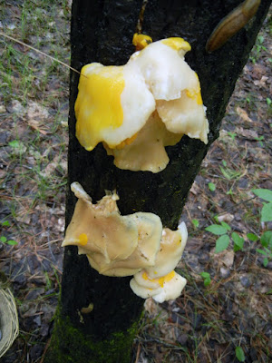
I found my first fungicolous myxomycete (a slime mold living on a fungus) two years ago in the height of the Pleurotus populinus (spring oyster) season. If I hadn’t been deep in the woods, I would probably have been able to see it from a kilometer away, it was such an unearthly bright yellow-orange. Up close, it was clearly an extensive slime mold plasmodium that was attempting to engulf two separate clusters of oyster mushrooms that were well past their frying-pan expiry date.
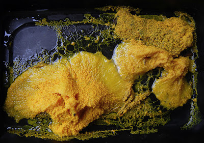
yellow plasmodium on mushroom
Badhamia utricularis plasmodium Day 1 (top) & Day 2 (bottom)

I could have left this spectacle where it was, but I’d been wanting a new pet, so I took the whole kit and caboodle home with me. I say “pet” because I’d read quite a bit about the plasmodium of a particular slime mold—Physarum polycephalum—and had been keeping my eyes peeled, hoping to find some that I could capture and tempt into solving mazes or planning efficient rail systems by baiting it with oatmeal as other people had been doing. It sounded like fun. Cheap fun.

The yellow plasmodium travelled on an off the mushrooms for several days.

First, though, I was curious about whether or not the plasmodium I’d found was actually consuming the oyster mushrooms I’d found it on, or if it was simply passing over them. I took photographs over several days and watched my myxo completely cover the Pleurotus, then move off into a corner for a few hours (perhaps to relax and digest?), then return to the quickly shrinking mushrooms and engulf them again. It repeated this pattern for four days, and during this time the mushrooms shrank and shrank and shrank. Unusually, there were no signs of the normal larvae that one finds gorging on the tender flesh of the most edible Pleurotus species, the bane of fungi-eating people everywhere, and I believe this was solely due to the active presence of the plasmodium, which had either deterred the inevitable laying of eggs by insects or had somehow made eggs that had been laid unviable.

Plasmodium Day 4 (top) and Day 5 (bottom)

An interesting thing about Physarum polycephalum, which is studied in labs around the world, is that there are very few pictures of mature sporangia—the structures that produce spores—on the internet. There are even fewer examples of a progression from plasmodium to sporangia. Despite this lack of sporangial evidence, there are many photographs of yellow plasmodium on line that look just like mine, and a great many of these are labeled as Physarum polycephalum. Some are engulfing mushrooms. Some of these mushrooms are species of Pleurotus. Furthermore, in a 1987 paper, Quentin D. Wheeler states that Pleurotus is “so often the substrate over which this slime mold grows that the plasmodium is a field identification “character” of the fungus itself (Lincoff 1981*).” Obviously, I had found my first Physarum polycephalum. Or had I?

slime mold sporangia beginning to form
The sporangia beginning to form.

From early on, I was a tad disturbed by the colour of my new “pet,” which seemed a little off, a little too orange-yellow compared to the yellow to greenish-yellow it was supposed to be if it was really Physarum polycephalum. 

Sporangia beginning to get their mature blueish grey colour.
And then my plasmodium crawled away from the flattened, liquefied remnants of the mushrooms it had been consuming and formed sporangia that were not at all what they were supposed to be. In fact, what they turned into was a big whack of Badhamia utricularis.

Badhamia utricularis sporangia are brownish  before drying to blue-grey.
Badhamia utricularis sporangia are brownish
before drying to blue-grey.

Surprisingly, this was not an anomaly. I found another batch of mature B. utricularis in the winter that had apparently been feeding on a Trametes before it, too, had formed sporangia. And then, again, this past spring, I found new clusters of Pleurotus populinus being eaten by, once again, B. utricularis at two different sites.
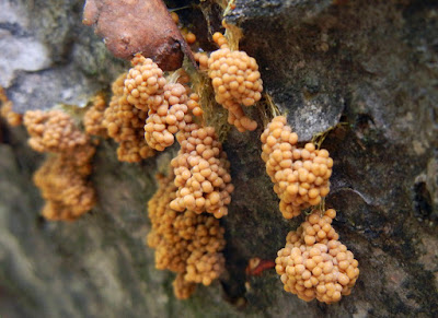
slime mold hangs in clusters by shrivelled yellowish strands.
B. utricularis sporangia are usually pendant, hanging
in clusters by shrivelled yellowish strands.

So I did a little more detective work and found a most entertaining 1906 article written by a past president of the British Mycological Society, Arthur Lister: “Notes on the Plasmodium of Badhamia utricularis and Brefeldia maxima.”

Arthur Lister, who kept slime moulds as pets.

Arthur Lister made his money as a wine merchant, then retired so he could devote his time and energies to the study of botany, primarily myxomycology, which allowed him time to observe and write notes about a pet B. utricularis that he “kept in constant streaming movement on various types of woody fungi for more than a year.” (Sometimes I wish I’d been a born in the 1800s. Male. To a family of means.) (Oh, yeah—with a daughter, Gulielma, who would do all the gorgeous, detailed illustrations for my book: A Monograph of the Mycetozoa (1894.))
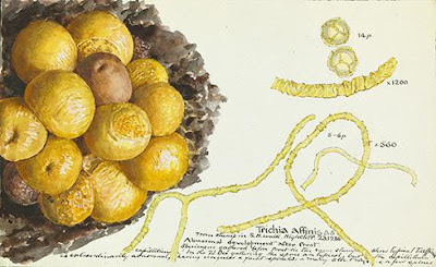

Gulielma Lister
Gulielma Lister's illustration of Trichia affinis.
Gulielma Lister and her amazing illustration of Trichia affinis

Lister found his Badhamia on a garden log where it was consuming portions of an extensive patch of Coniophora puteana. He captured some of it and kept it alive in specially constructed boxes, allowing it to feed on thin species of Daedalea, Bjerkadera adusta, Trametes versicolor, and, its particular favourite, Stereum hirsutum. It also nibbled on Armillaria mellea and Suillus grevillei, and partially consumed some Amanita rubescens. It was not thrilled, however, when offered Hypholoma fasciculare. Though it politely fed briefly on this mushroom, it did not agree with it at all and the plasmodium, which was clearly ailing, did not look itself again until it had been rejuvenated with a fresh piece of Stereum hirsutum.

I found this clutch in the winter.

During Lister’s year of experimentation with this plasmodium, numerous colonies made the change to sporangia. Others colonies, however, though being given exactly the same conditions as the ones that had matured, continued to stream and feed. Lister was unable to discover why some chose to form sporangia while others did not. And nor have I.
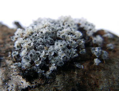

Weathered Badhamia utricularis

This spring’s batch of plasmodium that I brought home swarmed over the oyster mushrooms for several days until it had turned them into a pool of mushroom-shaped muck and then it just went pfft. Total dud. Though the plasmodium of B. utricularis is capable of forming dormant sclerotium that can be revived when moistened, my last plasmodium simply shriveled up and died.

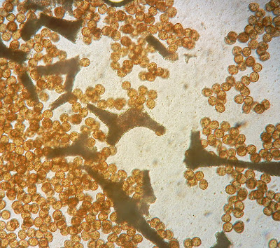
Badhamia utricularis spores & capillitium

According to Lister’s notes, he never offered his plasmodium pets any sort of Pleurotus. I wish he had so I could read his observations. In just two years, I’ve found it feeding on Pleurotus populinus three separate times, so obviously the organism takes some pleasure in eating it. I believe other people have found B. utricularis on Pleurotus as well, other people who have jumped on a Physarum polycephalus bandwagon for no other reason than their having found an extensive yellow plasmodium engulfing a mushroom or other fungus on a log. But I think it’s a fool’s game to try to name a plasmodium to species without having the proof of sporangia. Pick it up, I say. Take it home. Enjoy the antics of your new “pet” until it chooses to reveal who it really is!
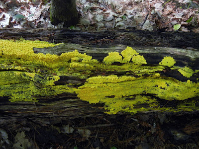

Fuligo septica plasmodium
A massive expanse of Fuligo septica plasmodium like this could be 
mistaken for either Physarum polycephalum or Badhamia utricularis.


References & Resources:


Lister, Arthur. Notes on the Plasmodium of Badhamiautricularis and Brefeldia maxima Annals of Botany, Volume 2 1906 

Wired magazine's article about Physarum polycephalum & Tokyo rail system

Scientific American's article about Physarum polycephalum and mazes

Quentin D. Wheeler, A New Species of Agathidium Associated with an "Epimycetic" Slime Mold Plasmodium on Pleurotus Fungi (Coleoptera: Leiodidae-Myxomycetes: Physarales-Basidiomycetes: Tricholomataceae). The Coleopterists Bulletin Vol. 41, No. 4 (Dec., 1987), pp. 395-403

*Lincoff, G. The Audubon Society Field Guide to North American Mushrooms, 1981 – In Lincoff’s defence, he does not say exactly what Wheeler says in the Audubon Guide. What he says in the entry for Physarum polycephalum is that they can be found “on dead wood and fleshy fungi,” and, under the entry for Pleurotus ostreatus: “It is sometimes covered by the yellow early stage of a slime mold.”

This paper is “based on greenish yellow plasmodium collected on pilei of Pleurotus sapidus,” material that was cultured and proved to be P. polycephalum:
Howard, F. L., The Life History of Physarum polycephalum. American Journal of Botany Vol. 18, No. 2 (Feb., 1931), pp. 116-133


Wednesday 27 May 2015

"Very Strange Conidia": Phragmotrichum thornhilliae

Phragmotrichum chailletii appears on spruce cones in early spring.

Here’s a not-terribly-exciting-looking little ascomycete that—if you live in northern areas of Europe or North America—you’ve probably stepped on in the woods without even knowing it. Phragmotrichum chailletii forms tiny (0.5-1 mm) black pustules, or perithecia, that erupt from the upper surface of the scales of fallen spruce cones. It frequently grows in dense numbers on the scales, though the ones I’ve found have always been scattered. I’ve found it two years in a row, both times very close to the same date, once on April 21, and once on April 24. It seems to like snowmelt moisture and cold temperatures.


Phragmotrichum chailletii is little studied, so there’s not much known about it other than that it’s apparently not a pathogen of spruce trees since it’s only found on fallen spruce cones, usually in early to late spring. It also only produces conidia, or asexual spores; a teleomorph, or "perfect" form of this fungus that produces sexual spores, is unknown. 

Phragmotrichum chailletii conidia

These conidia are what’s really fun about P. chailletii. In the original description (Kunze & Schmidt, 1923 - see reference below), the shape and structure of these conidia are described as "very strange" with no known analogy in mycology. 


The conidia develop in chains.

First off, they’re quite large—25-45 µm—which makes them photogenic. They’re brown and and multi-septate—“mûriforme” in French, or shaped like a blackberry. They’re produced in chains, that, when you’re lucky, show the progression of development from amorphous, non-septate tubes, to single septate, to double, and so on, until to they’re fully developed multi-septate “blackberries.” It gives me joy to look at them, as if I’m looking at a long-lost M. C. Escher print.

Conidia are released from the fruiting body of Phragmotrichum
chailletii
when it splits open. (click to enlarge)
This is a view that reminded me of M.C. Escher's work.
I almost expected to see lizards
.


References

Original description (in German): Kunze & J.C. Schmidt 1823, Mykol. Hefte 2: 84, t. 2:4


This paper offers a key to four Phragmotrichum species: B. Sutton, D.K. Sandhu, Phragmotrichum pini (W.B. Cooke), Transactions British Mycological Society, 52 (I), 67-71 (1969)

Tuesday 28 April 2015

A Teratological Mystery from 1744: Agaricus ramosus/Boletus ramoso/Boletus rangiferinus/Polyporus squamosus

Ehret Agaricus ramosus or abnormal growth of Polyporus squamosus
Georg Dionysius Ehret's 1744 illustration
of "Agaricus ramosus"
I was leisurely poking around the Wellcome Library's astonishing collection of hi-res historical images when I was stopped in my tracks by this fabulous, otherworldly image of a fungus. What the heck is it supposed to be? 

My first thought was that it was either a poor rendering, or something invented. But the man who made this hand-painted engraving was no slouch. In fact, he was one of the most celebrated botanical illustrators of his time: Georg Dionysius Ehret. Born in Germany in 1708, Ehret began his career as a gardener at a local castle. But he also studied and drew plants and insects with enough detail that before he was thirty he was already collaborating in Holland with Carl Linnaeus in the production of the important botanical work, Hortus Cliffortianus. Eventually he moved to London where he specialized in illustrating the numerous exotic plants that were regularly being brought to England from the colonies. He was lauded not just for his style, but also for his anatomical accuracy in rendering these plants.


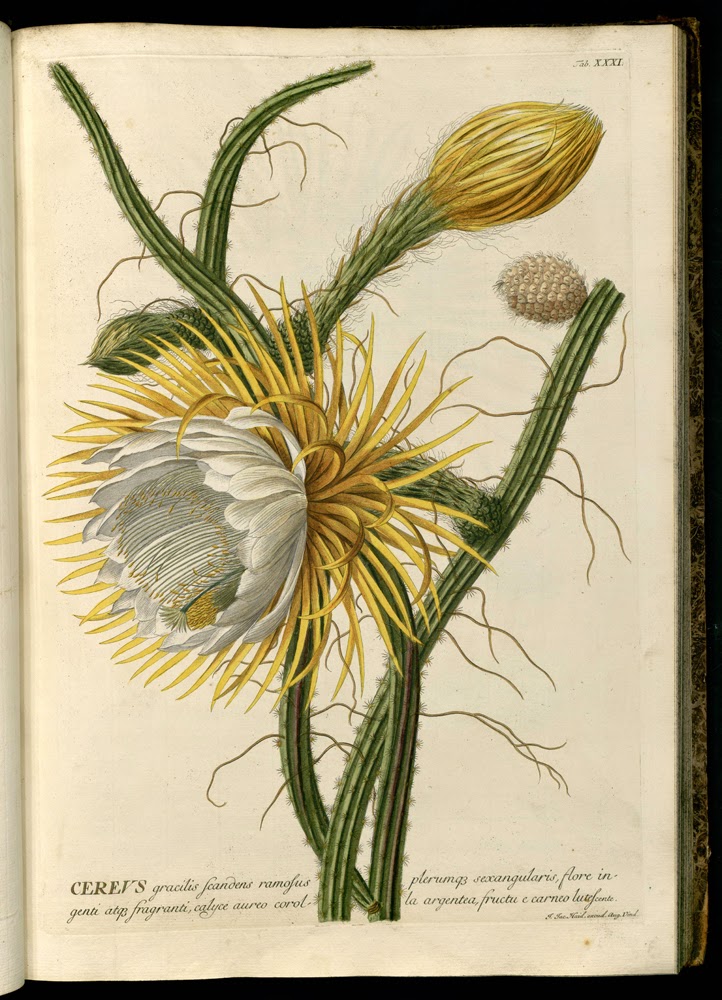
Ehret Selenicereus gradiflorus 1750 from Plantae selectae
Ehret's 1750  illustration of Selenicereus
grandiflorus
from Plantae selectae
Though he rarely depicted fungi, it's obvious that Ehret knew what he is doing. Which means we have to assume that the illustration of the antlered fungus must be a fairly faithful rendering of what was found growing in Mr. Winckles’ cellar.


Helvella mitra water-colour by Ehret
Ehret's 1760 watercolour of "Helvella mitra"—a rare, for him, rendering of a fungus.

But What the Heck Is It?


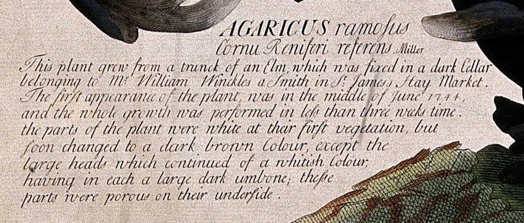
Ehret's description of Agaricus ramous Ployporus squamosus
Close-up of Ehret's description of Agaricus ramosus
Though called "Agaricus ramosus," the fungus depicted is clearly not an Agaricus of any kind, certainly not as we now know the genus; Agaricus species all have gills, and Ehret clearly states—and shows—that this “plant” has pores. Of course, 1744 was early days in terms of fungal taxonomy and, at the time, the name Agaricus was widely applied to many fungi, including polypores. 

With no other clues, I Googled "Agaricus ramosus" to see what would happen. The first page of hits turned up nothing other than reproductions of the same illustration—no help at all. Mycobank was also no help, nor, surprisingly, was Index Fungorum, the international index of formal fungi names. I plowed on and eventually found reference to Ehret's fungus in a 1745 issue of The Royal Society’s Philosophical Transactions, a copy of which, despite being 270 years old, was easy to find (I can't tell you how much I love the World Wide Web!):

"An Account of a New Species of Fungus by John Martyn [professor of botany, Cambridge] communicated in a Letter to the President:

“In the latter Part of the Summer of the Year 1744, Mr Ehret the Painter brought me a Fungus of a very extraordinary Shape and Size, which had been found growing on a Piece of the Trunk of an Elm, in a damp Cellar in the Hay-Market.

“The whole Plant was about two Feet in Height; and, at first Sight, seemed not very unlike the Horns of some Deer, being variously branched, and covered with a thick Down. It was of a spongeous Substance, and of a dusky-red Colour inclining to Black. The Tips of the smaller Branches were of a Cream-Colour. The larger Branches or rather the Tops of the whole Plant, were expanded in Form of a Funnel, smooth on the concave, and full of Pores on the convex Side. The inner and lower Part of the Funnel was of the same Colour with the Stalk; the rest of it was of a cream-Colour.

“I have not been able to find, that this Plant has been mentioned by any Author: and I am persuaded, that it is a new Species; and, perhaps, the remarkable branching of the Stalks may induce some to think it a new Genus..." 

Martyn, however, goes on to decide that it is not a new genus, and, because its cap is not lamellated, he makes it a Boletus, settling on the name Boletus ramoso—a name that goes absolutely nowhere on the internet.

But then I discover that Ehret’s image had been listed in a library catalogue as Boletus rangiferinus, a name James Bolton came up with in 1888 after being shown what he described as a 

"curious and extraordinary fungus growing on a Log of Wood, in the cellar of a Publick House in Leeds, in the beginning of October...I had an opportunity of drawing it when fresh and newly gathered, but it is in the possession of a Man, of such a temper (who is no Naturalist) that no offers I could make him would prevail him to part with it!" 

I typed Botetus rangiferinus into Google...


...and Suddenly Everything Falls into Place


It turns out that this bizarre antlered fungus that grows in dark, dank cellars, descriptively named Agaricus ramous and Boletus ramosa and Boletus rangiferinus (ramose=branched and rangifer=reindeer), is none other than—drum roll, please—an abnormal, or teratological form of the extremely common, cosmopolitan wood decayer, Polyporus squamosus, aka Dryad's Saddle (a name that goes back hundreds of years) or Pheasant's Back (a 20th century name). Whatever you prefer to call it, P. squamosus apparently does very peculiar things in the dark. 


Edible Polyporus squamosus, Pheasant back, Dryad's saddle
Perfectly normal Polyporus squamosus growing on a standing dead elm
There are other descriptions of this "cellar fungus" "monstrosity" to be found on the internet. According to an article in the The Midland Naturalist (1888), one was found in a wine cellar with a 

"dark coloured base about six inches wide, and from this arose a number of clavaria-like processes, some of which were branched. These clavaria-like branches were from three to six inches long, and were of a pale fawn colour." 

Another, smaller example, was found growing in the rafters of a house when a floor was removed for repairs.

And then I found another illustration of the antler deformity, this one by James Sowerby in English Fungi or Mushrooms (1797). That sure looks like an antler to me.


mutant mushroom Polyporus squamosus  James Sowerby, English Fungi or Mushrooms (1797)
A clearly depicted Polyporus squamosus "antler" by
James  Sowerby in English Fungi or Mushrooms (1797)
One can hardly blame those who saw these "antler" growths to think they'd stumbled across an unnamed species. I would probably have thought the same thing, particularly since the first time I found baby P. squamosus—having only seen large mature ones before—I initially didn't have a clue what they were...and they weren't even sporting black antlers. But I'd like to see the "clavaria-like" form, so I have a mind to take a chainsaw out back and cut a piece off a productive dead elm to put in my root cellar to see if I can grow my own teratological P. squamosus formation. 

Young Dryad's saddle, pheasant's back, Polyporus squamosus
Polyporus squamosus "buttons" can be confusing to the uninitiated.


References

Georg Dionysius Ehret on Wikipedia
An Account of a New Species of Fungus by John Martyn, The Royal Society’s Philosophical Transactions 1745
W. B. Grove. A Cellar Fungus,The Midland Naturalist (1888)
"Mutation Collection" of fungi on Mushroom Observer

Polyporus squamosus on the web:

First Nature
Some cooking tips on Mushroom-collecting.com